Shade-Matching Challenge
A Single Central Incisor
Achieving a good color match when restoring a single incisor is probably among the most difficult aesthetic challenges for any dentist (Figure 1). While the latest technology can be found in most modern dental offices such as CBCT; laser; CAD/CAM; and less common, the more expansive spectrophotometric instruments;1 the vast majority of clinicians still conduct dental shade selection by using a nearby window for a natural light source or, if they are fortunate, pass the buck by simply sending the patient to the dental laboratory technician to take and map the shade. There must be a better way and, in the authors’ opinions and experience, there is! A simple and inexpensive handheld portable LED light source, the Rite-Lite 2 HI CRI Shade Matching Light (AdDent), is now available help achieve an excellent restorative shade match.2-4
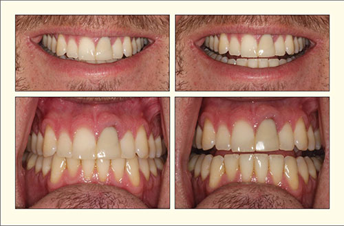
Figure 1. Pre-op photos of mismatched crown on nonvital central incisor with gingival inflammation.
DESCRIBING A SHADE
Shade matching is an interdisciplinary process that requires the clinician to communicate with the dental laboratory team using a common language and images (shade-mapping and photographs). Thus, shade matching relies on perception and interpretation of the evidence.
Color, commonly referred to as the shade, is divided into 3 components.
- Hue refers to the basic color (eg, red, blue, green).
- Chroma refers to the intensity of the color (eg, fire-truck red versus pastel pink).
- Value refers to the brightness of the color (eg, the range of gray from black to white).
All these components should not be overlooked, or else a wrong interpretation of color may lead to an undesired result. For example, how often have you told your ceramist to make the cuspids slightly darker when restoring an anterior case? However, your real intent was to make the cuspids warmer with more chroma but not darker (lower value).
It is important to realize that the correct language helps in the interpretation of the evidence. Acquiring the evidence relies on the physiology of our eyes and the transmitted light.5
How We Perceive Color
We perceive color using cone cells that are located in the fovea in the middle of the retina. Cone cells are few in numbers and are divided into 3 groups. Each group responds to a specific color: red, blue, or green.6 Cone cells fatigue extremely fast, since they are limited in number. For example, if you stare at a color, such as red lipstick, the red cone cells will shut down after 30 seconds. This will leave you seeing only the combination of colors provided by the green and blue cells. This is why it is necessary to create a neutral background for your eyes before selecting a shade. Ideally, the walls in the room should be gray or white. Ask your female patients to remove their lipstick and place a pale blue or grey bib over their clothes.7
View Full Article